Mouse models are commonly used for studying hepatocellular carcinoma (HCC) biology and exploring new therapeutic interventions. Currently three main modalities of HCC mouse models have been extensively employed in pre-clinical studies including chemically induced, transgenic and transplantation models. Among them, transplantation models are preferred for evaluating in vivo drug efficacy in pre-clinical settings given the short latency, uniformity in size and close resemblance to tumors in patients. However methods used for establishing orthotopic HCC transplantation mouse models are diverse and fragmentized without a comprehensive comparison. Here, we systemically evaluate four different approaches commonly used to establish HCC mice in preclinical studies, including intravenous, intrasplenic, intrahepatic inoculation of tumor cells and intrahepatic tissue implantation. Four parameters--the latency period, take rates, pathological features and metastatic rates--were evaluated side-by-side. 100% take rates were achieved in liver with intrahepatic, intrasplenic inoculation of tumor cells and intrahepatic tissue implantation. In contrast, no tumor in liver was observed with intravenous injection of tumor cells. Intrahepatic tissue implantation resulted in the shortest latency with 0.5 cm (longitudinal diameter) tumors found in liver two weeks after implantation, compared to 0.1cm for intrahepatic inoculation of tumor cells. Approximately 0.1cm tumors were only visible at 4 weeks after intrasplenic inoculation. Uniform, focal and solitary tumors were formed with intrahepatic tissue implantation whereas multinodular, dispersed and non-uniform tumors produced with intrahepatic and intrasplenic inoculation of tumor cells. Notably, metastasis became visible in liver, peritoneum and mesenterium at 3 weeks post-implantation, and lung metastasis was visible after 7 weeks. T cell infiltration was evident in tumors, resembling the situation in HCC patients. Our study demonstrated that orthotopic HCC mouse models established via intrahepatic tissue implantation authentically reflect clinical manifestations in HCC patients pathologically and immunologically, suggesting intrahepatic tissue implantation is a preferable approach for establishing orthotopic HCC mouse models.
文章引用产品列表
-
- EK210 486 Citations
- FEATURED ELISA KIT, ELISA试剂盒
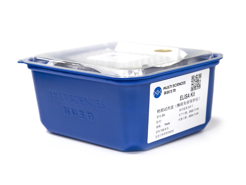
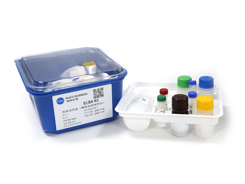
Mouse IL-10 ELISA Kit检测试剂盒(酶联免疫吸附法)
- ¥1,600.00 – ¥10,800.00


