Introduction:Rheumatoid arthritis (RA) is a chronic inflammatory disease involving a variety of immune cells, including adaptive T and B cells and innate lymphoid cells (ILCs). Understanding the pathogenic role of these immune cells in RA provides new insights into the intervention and treatment of RA.
Methods:A total of 86 patients with RA (RA group) and 50 healthy controls (HC) were included in the study. The immune cells of CD4+, CD19+ B, NK, Th17, Treg, ILCs, and their subsets (i.e., ILC1s, ILC2s, and ILC3s) were characterized in peripheral blood mononuclear cells by flow cytometry. Cytokines (i.e., IFN-γ, IL-4, IL-10, IL-17A, IL-22, and IL-33) in sera were detected using ELISA. The above immune cells and cytokines were analyzed in patients with different disease activity status and positive ( +) or negative ( -) rheumatoid factor (RF)/anti-citrullinated protein antibodies (ACPA).
Results:Patients with RA had higher percentages of CD4+ T, CD19+ B, Th17, ILC2s, and ILC3s and lower percentages of Treg and ILC1s than HC. Patients with RA had elevated levels of IFN-γ, IL-4, IL-17A, and IL-22 and decreased level of IL-10. Compared with HC, patients with high disease activity had higher percentages of Th17, ILC2s, and ILC3s; lower percentages of ILC1s; and lower level of IL-10. The percentage of Treg cells in remission, low, moderate, and high disease activities decreased, whereas the level of IL-17A increased compared with HC. Furthermore, RF+ or ACPA+ patients exhibited elevated percentages of CD19+ B, ILC2s, and ILC3s and had decreased percentage of ILC1s and Treg cells than HC. The percentage of Th17 cells increased in RF-/ACPA- and RF+/ACPA+ patients. However, the above immune cells between RF or ACPA positive and negative patients were not significantly different.
Conclusion:Th17, Treg, and ILC subset dysregulations are present in patients with RA but may not be associated with conventionally defined seropositive RF and ACPA. Key Points • Th17, Treg, and ILC subset dysregulations are present in patients with RA but may reflect inflammation rather than specific diseases and stages. • No difference for the distribution of Th17, Treg, and ILC subsets between RF+ and RF- patients and between ACPA+ and ACPA- patients. The screening spectrum of RF and ACPA serology should be expanded to elucidate the role of immune cells in RA pathogenesis.
文章引用产品列表
-
- EK180 169 Citations
- ELISA试剂盒
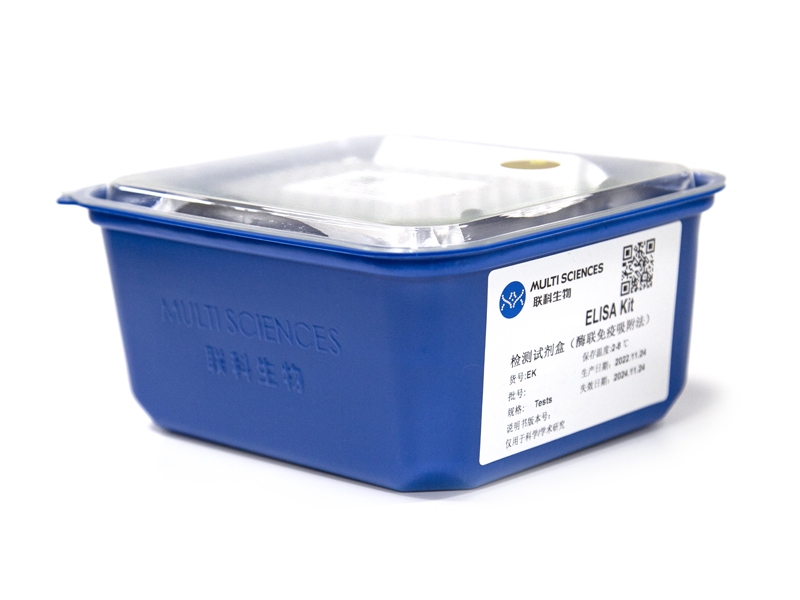
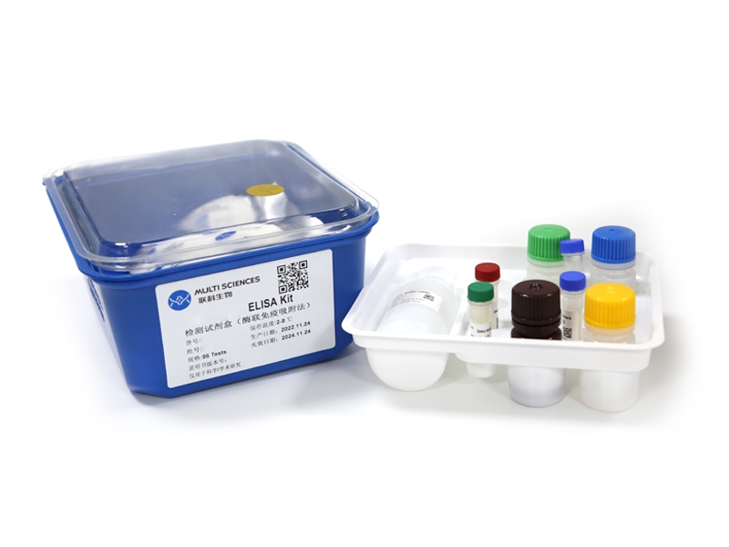
Human IFN-gamma ELISA Kit检测试剂盒(酶联免疫吸附法)
- ¥1,600.00 – ¥10,800.00
-
- EK180HS 130 Citations
- 高敏试剂盒
Human IFN-γ High Sensitivity ELISA Kit检测试剂盒(酶联免疫吸附法)
- ¥2,000.00 – ¥3,400.00
-
- EK133 12 Citations
- ELISA试剂盒
Human IL-33 ELISA Kit检测试剂盒(酶联免疫吸附法)
- ¥1,600.00 – ¥2,650.00
-
- EK133HS 10 Citations
- 高敏试剂盒
Human IL-33 High Sensitivity ELISA Kit检测试剂盒(酶联免疫吸附法)
- ¥2,000.00 – ¥3,400.00
-
- EK122HS 7 Citations
- 高敏试剂盒
Human IL-22 High Sensitivity ELISA Kit检测试剂盒(酶联免疫吸附法)
- ¥2,000.00 – ¥3,400.00
-
- EK122 7 Citations
- ELISA试剂盒
Human IL-22 ELISA Kit检测试剂盒(酶联免疫吸附法)
- ¥1,600.00 – ¥2,650.00
-
- EK117HS 46 Citations
- 高敏试剂盒
Human IL-17A High Sensitivity ELISA Kit检测试剂盒(酶联免疫吸附法)
- ¥2,000.00 – ¥3,400.00
-
- EK117 52 Citations
- ELISA试剂盒
Human IL-17A ELISA Kit检测试剂盒(酶联免疫吸附法)
- ¥1,600.00 – ¥2,650.00
-
- EK110HS 115 Citations
- 高敏试剂盒
Human IL-10 High Sensitivity ELISA Kit检测试剂盒(酶联免疫吸附法)
- ¥2,000.00 – ¥3,400.00
-
- EK110 138 Citations
- ELISA试剂盒
Human IL-10 ELISA Kit检测试剂盒(酶联免疫吸附法)
- ¥1,600.00 – ¥2,650.00
-
- EK104HS 45 Citations
- 高敏试剂盒
Human IL-4 High Sensitivity ELISA Kit检测试剂盒(酶联免疫吸附法)
- ¥2,000.00 – ¥3,400.00
-
- EK104 61 Citations
- ELISA试剂盒
Human IL-4 ELISA Kit检测试剂盒(酶联免疫吸附法)
- ¥1,600.00 – ¥2,650.00


