Background Studying the potential etiology and pathogenesis of tuberculosis-associated chronic obstructive pulmonary disease (TOPD) from an autoimmunity perspective may provide insights into peripheral blood autoantibodies and immune cells, as well as their interactions.Methods This study examined the serum autoantibody repertoire in healthy individuals, patients with chronic obstructive pulmonary disease (COPD), patients with pulmonary tuberculosis (TB), and TOPD patients using the HuProtTM protein chip. Autoantigens in the peripheral blood of TOPD patients were verified using ELISA assay. Various epitopes and immune simulation were predicted using bioinformatic methods. Flow cytometry was employed to detect macrophages(Mφ), T cells, and innate lymphoid cells (ILCs) in the peripheral blood.Results COPD patients displayed distinct alterations in their IgG and IgM autoantibodies compared to the other groups. GeneOntology (GO) and Kyoto Encyclopedia of Genes and Genomes(KEGG)analyses revealed that these autoantibodies were associated with regulating macrophages, T cells, and B cells. ELISA results confirmed the upregulation of expression of proliferating cell nuclear antigen (PCNA), Mitogen-Activated Protein Kinase 3 antigen (MAPK3), and threonine protein kinase 1 antigen (AKT1) proteins in the peripheral blood of TOPD patients. Bioinformatic analysis predicted multiple potential epitopes in Th, CTL, and B cells. Immune simulation results demonstrated that PCNA, MAPK3, and AKT1 can activate innate and adaptive immune responses and induce the expression of different cytokines, such as IFN-g and IL-2. Furthermore, data obtained from flow cytometry assay revealed an upregulation in the face of Th1 cells in the peripheral blood of TOPD patients.Conclusion Tuberculosis infection can effectively induce autoimmune responses, contributing to increased expression of Th1 cells and associated cytokines, ultimately leading to immune dysregulation. Furthermore, the accumulation of pulmonary inflammatory response facilitates the progression of TOPD and is helpful for the clinical diagnosis and the development of targeted therapeutic drugs for this disease.
文章引用产品列表
-
- EK180 169 Citations
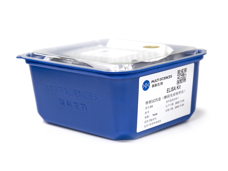
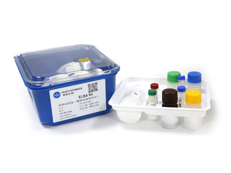
- ELISA试剂盒
Human IFN-gamma ELISA Kit检测试剂盒(酶联免疫吸附法)
- ¥1,600.00 – ¥10,800.00
-
- EK180HS 130 Citations
- 高敏试剂盒
Human IFN-γ High Sensitivity ELISA Kit检测试剂盒(酶联免疫吸附法)
- ¥2,000.00 – ¥3,400.00
-
- EK117HS 46 Citations
- 高敏试剂盒
Human IL-17A High Sensitivity ELISA Kit检测试剂盒(酶联免疫吸附法)
- ¥2,000.00 – ¥3,400.00
-
- EK117 52 Citations
- ELISA试剂盒
Human IL-17A ELISA Kit检测试剂盒(酶联免疫吸附法)
- ¥1,600.00 – ¥2,650.00
-
- EK110HS 115 Citations
- 高敏试剂盒
Human IL-10 High Sensitivity ELISA Kit检测试剂盒(酶联免疫吸附法)
- ¥2,000.00 – ¥3,400.00
-
- EK110 138 Citations
- ELISA试剂盒
Human IL-10 ELISA Kit检测试剂盒(酶联免疫吸附法)
- ¥1,600.00 – ¥2,650.00
-
- EK104HS 45 Citations
- 高敏试剂盒
Human IL-4 High Sensitivity ELISA Kit检测试剂盒(酶联免疫吸附法)
- ¥2,000.00 – ¥3,400.00
-
- EK104 61 Citations
- ELISA试剂盒
Human IL-4 ELISA Kit检测试剂盒(酶联免疫吸附法)
- ¥1,600.00 – ¥2,650.00


